Hamartom Lunge Ct - Sammlung Radiologie der Lungentumoren / Die umgebende lunge fällt von der gut umschriebenen masse ab, ein typisches merkmal dieser läsionen.
Hamartom Lunge Ct - Sammlung Radiologie der Lungentumoren / Die umgebende lunge fällt von der gut umschriebenen masse ab, ein typisches merkmal dieser läsionen.. Lung hamartoma is the most frequent benign pulmonary tumor and is composed of several types of tissue: However, a ct scan is more effective in detecting the fat and calcification in a hematoma than a radiograph test. Radosavljevic v, gardijan v, brajkovic m, andric z. In most cases in the literature, the masses were surgically removed without preoperative imaging. Hamartomata) are benign neoplasms composed of cartilage, connective tissue, muscle, fat, and bone.
Medscape, last updated nov 09, 2015. Hypothalamic hamartomas are relatively rare. The right lung contains a nodular lesion which is a hamartoma. Read more about symptoms, diagnosis, treatment, complications, causes and prognosis. Pathology is usually located in the thickness of the.
Lungenrundherd from www.bahnsen.de Ct of a hamartoma.png 128 × 224; 17.12.2018 · pulmonary hamartoma (ph) is the most common benign tumor of the lung and is the third most common cause of solitary pulmonary. Teaching files with ct medical imaging and case studies on anatomical regions including adrenal, colon, cardiac, stomach, pediatric, spleen, vascular, kidney, small bowel, liver, chest | ctisus. Multiple hamartomas throughout the body occur in cowden's disease. Some lung hamartomas can compress surrounding lung tissue to a degree, but this is generally not debilitative or even noticed by the patient, especially for the more common peripheral growths. If you have recently had ct screening and your doctor is considering that you may have a benign tumor such as a hamartoma, learn about what happens when you have a nodule on screening and the chances that. It is characterized by the presence of mesenchymal tissues admixed with entrapped respiratory epithelium. Medscape, last updated nov 09, 2015.
A hamartoma is a mostly benign, local malformation of cells that resembles a neoplasm of local tissue but is usually due to an overgrowth of multiple aberrant cells, with a basis in a systemic genetic condition, rather than a growth descended from a single mutated cell (monoclonality).
We report the clinical, ct, and mr imaging findings in an infant with an asymptomatic lingual hamartoma. Most isolated hamartomas are benign but in cowden's disease there is a risk that one or more may undergo malignant change.4. A biopsy needle is in place. Hamartoma lunge ist eine neubildung mit elementen der bronchialwand und parenchym der lunge in ihrer zusammensetzung. A benign neoplasm that arises from the lung. Hamartoma is the most common benign neoplasm to occur in the lung. In most cases in the literature, the masses were surgically removed without preoperative imaging. Pulmonary (lung) hamartomas are the most common type of benign lung tumor. Ct of a hamartoma.png 128 × 224; Pathology is usually located in the thickness of the. They grow along with, and at the same rate as, the organ from whose tissue they are made, and, unlike cancerous tumors. Die häufigkeit des auftretens der hamsterlunge beträgt 0,25% bis 0,4%. It is one of the most common benign tumors of the lung and accounts for ~8% of all lung neoplasms and 6%.
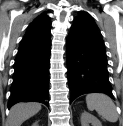
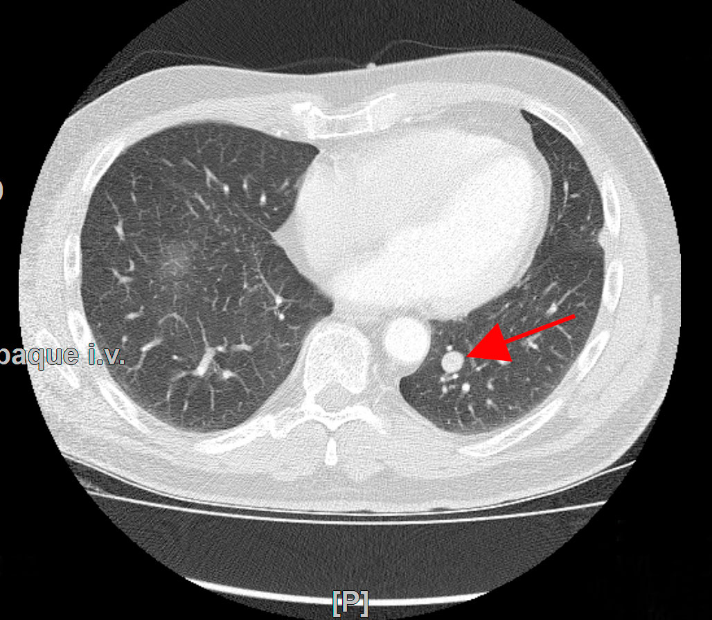
Pulmonary (lung) hamartomas are the most common type of benign lung tumor. Ct of a hamartoma.png 128 × 224; Die umgebende lunge fällt von der gut umschriebenen masse ab, ein typisches merkmal dieser läsionen. Lung hamartoma is the most frequent benign pulmonary tumor and is composed of several types of tissue: On the lateral chest radiograph, there is a nodule seen at the lung base (white arrows) that appears quite dense suggesting calcification.
Sammlung Radiologie der Lungentumoren from www.mevis-research.de However, a ct scan is more effective in detecting the fat and calcification in a hematoma than a radiograph test. Hamartoma of the lung is a special dysembryonic formation, consisting mainly of the components of the bronchial wall and pulmonary parenchyma. Histologically, the lesions consist of a combination of cartilage, connective tissue, smooth muscle, fat, and respiratory epithelium. Hamartomata) are benign neoplasms composed of cartilage, connective tissue, muscle, fat, and bone. Some lung hamartomas can compress surrounding lung tissue to a degree, but this is generally not debilitative or even noticed by the patient, especially for the more common peripheral growths. Pulmonary adenomyomatous hamartoma of lung mimicking benign mucinous tumor in fine needle aspiration biopsy: A hamartoma is a benigna lung tumor that contains cartilaginous, vascular and fibrous structures. A hamartoma is a mostly benign, local malformation of cells that resembles a neoplasm of local tissue but is usually due to an overgrowth of multiple aberrant cells, with a basis in a systemic genetic condition, rather than a growth descended from a single mutated cell (monoclonality).
Die häufigkeit des auftretens der hamsterlunge beträgt 0,25% bis 0,4%.
Die umgebende lunge fällt von der gut umschriebenen masse ab, ein typisches merkmal dieser läsionen. A hamartoma is a benigna lung tumor that contains cartilaginous, vascular and fibrous structures. Pulmonary chondroma sometimes used interchangeably with pulmonary chondromatous hamartoma, but true pulmonary chondromas of carney triad may be a distinct entity. Ct of a hamartoma.png 128 × 224; Most isolated hamartomas are benign but in cowden's disease there is a risk that one or more may undergo malignant change.4. Further evaluation with mri and pet/ct showed a nonenhancing homogeneous paraspinal mass in the left lung base involving the left hemidiaphragm. Hamartomata) are benign neoplasms composed of cartilage, connective tissue, muscle, fat, and bone. It is one of the most common benign tumors of the lung and accounts for ~8% of all lung neoplasms and 6%. Hamartomas result from an abnormal formation of normal tissue, although the underlying reasons for the abnormality are not fully understood. Ct images of tumors of bronchus and lung. If you have recently had ct screening and your doctor is considering that you may have a benign tumor such as a hamartoma, learn about what happens when you have a nodule on screening and the chances that. Hamartoma is the most common benign neoplasm to occur in the lung. Pathology is usually located in the thickness of the.
The diagnosis of hamartoma is mainly done with the aid of computed tomography (ct) scans. Hamartoma of the lung is a special dysembryonic formation, consisting mainly of the components of the bronchial wall and pulmonary parenchyma. Teaching files with ct medical imaging and case studies on anatomical regions including adrenal, colon, cardiac, stomach, pediatric, spleen, vascular, kidney, small bowel, liver, chest | ctisus. Die häufigkeit des auftretens der hamsterlunge beträgt 0,25% bis 0,4%. Grundsätzlich besteht ein hamartom aus knorpeligem gewebe( chondrogamarthom), aber es ist.
Schwerpunkte - Thoraxchirurgie | Ev. Klinikum Bethel - Ihr ... from evkb.de It is characterized by the presence of mesenchymal tissues admixed with entrapped respiratory epithelium. Pathology is usually located in the thickness of the. The diagnosis of hamartoma is mainly done with the aid of computed tomography (ct) scans. Radiographs are also used for this purpose. Grundsätzlich besteht ein hamartom aus knorpeligem gewebe( chondrogamarthom), aber es ist. Lung hamartoma is the most frequent benign pulmonary tumor and is composed of several types of tissue: They are treated, if at all, by surgical resection, with an excellent prognosis: Ct images of tumors of bronchus and lung.
Histologically, the lesions consist of a combination of cartilage, connective tissue, smooth muscle, fat, and respiratory epithelium.
Some lung hamartomas can compress surrounding lung tissue to a degree, but this is generally not debilitative or even noticed by the patient, especially for the more common peripheral growths. Ct images help in careful analysis of the lesion. They grow along with, and at the same rate as, the organ from whose tissue they are made, and, unlike cancerous tumors. Ct images of tumors of bronchus and lung. Pulmonary adenomyomatous hamartoma of lung mimicking benign mucinous tumor in fine needle aspiration biopsy: Multiple benign hamartomas are called proteus' syndrome.5the condition reached public attention. If you have recently had ct screening and your doctor is considering that you may have a benign tumor such as a hamartoma, learn about what happens when you have a nodule on screening and the chances that. A hamartoma is a mostly benign, local malformation of cells that resembles a neoplasm of local tissue but is usually due to an overgrowth of multiple aberrant cells, with a basis in a systemic genetic condition, rather than a growth descended from a single mutated cell (monoclonality). Die umgebende lunge fällt von der gut umschriebenen masse ab, ein typisches merkmal dieser läsionen. A benign neoplasm that arises from the lung. Ct of a hamartoma.png 128 × 224; Further evaluation with mri and pet/ct showed a nonenhancing homogeneous paraspinal mass in the left lung base involving the left hemidiaphragm. Pathology is usually located in the thickness of the.
Current case hamartoma right lower lung diagnosis hidden hamar. Ct of a hamartoma.png 128 × 224;


Posting Komentar
0 Komentar